- Lentivirus
- Adeno-Associated
- Adenovirus
- Pseudovirus
- Vector
- Synthesis
- Autophagy Research
- CRISPR/Cas9
- Noncoding RNA
- Luciferase Assay
- Reagents
WHAT ARE YOU LOOKING FOR?
Chaperone-mediated autophagy (CMA) mainly recognizes a special motif on the protein sequence through molecular chaperones, and transports the protein into lysosomes for degradation. CMA can degrade damaged or mutated proteins and is important for maintaining intracellular protein homeostasis. Can also degrade key proteins in lipid and glucose metabolism, cell cycle, DNA repair, and other processes, playing a regulatory role. Typically, some degree of CMA activity can be detected in almost all different cell types in tissues or cultures such as liver, kidney, brain, etc., and cellular stress maximizes activation of this pathway. Factors such as starvation, oxidants, pro-oxidants, and protein denaturing toxins all cause CMA activation. CMA has gradually become a hot topic in autophagy research as it can precisely regulate cellular activities by selectively recognizing and degrading certain proteins in cellular processes. There is also a strong correlation between CMA and major diseases such as metabolic disorders, neurodegeneration, immunodeficiency, diabetes, and cancer. The efficient CMA activity monitoring tool can provide great convenience for the study of its potential function in diseases.
| Tool | Function introduction | Provide products |
KFERQ-PAmCherry1 | Detection of CMA activity | LV/Ad/AAV |
PAmCherry1-KFERQ-NE |
*Note: LV: Lentivirus, Ad: Adenovirus, AAV: Adeno-associated virus.
KFERQ-PAmCherry1 and PAmCherry1-KFERQ-NE virus tools
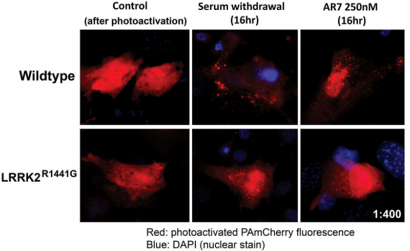
PAmCherry1 is a light-activated protein, which is lightless and needs to be activated by exposure to light at 405 nm for 5-10 minutes before it can be activated to emit red fluorescence (excitation 564 nm, emission 595 nm). The use of such fluorescent proteins reduces the effect of highly autofluorescent or cumulative autofluorescent pigment deposits on cellular observation. KFERQ-PAmCherry1 fuses to the photoactivated protein PAmCherry1 with the aid of the molecular chaperone recognition substrate motif KFERQ, which is converted into a CMA substrate, and when targeted to the lysosome, CMA activity can be detected in living cells by observing the number of red fluorescent spots.
Note: "NE" is a protein tag that can be used for protein expression detection.


MID: 30983487 (PAmCherry1-KFERQ-NE tool)

Contact Hanbio and leave your requirements. We will reply as soon as possible.