
- Lentivirus
- Adeno-Associated
- Adenovirus
- Pseudovirus
- Vector
- Synthesis
- Autophagy Research
- CRISPR/Cas9
- Noncoding RNA
- Luciferase Assay
- Reagents
WHAT ARE YOU LOOKING FOR?
01
Journal: Nature
IF: 64.8
Title: Tumour circular RNAs elicit anti-tumour immunity by encoding cryptic peptides
Method: Immunoblotting for Flag expression in HEK293T cells transfected with P-circ vector carrying an expression cassette for circFAM53B with a 3×Flag-coding sequence. P-circ vector was produced by cloning the circRNA sequence into the lentiviral vector (pHBLV-CMV-Circ-MCS-EF1-zsgreen-T2A-puro).
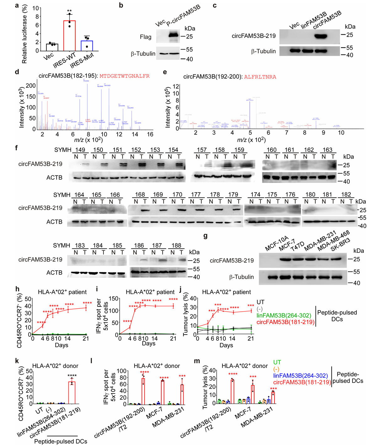
Fig. 1
02
Journal: Signal Transduction and Targeted Therapy
IF: 39.3
Title: Chronic pulmonary bacterial infection facilitates breast cancer lung metastasis by recruiting tumor-promoting MHCIIhi neutrophils
Method: The mouse breast carcinoma cell Line 4T1 was transduced with firefly luciferase lentiviral expression particles (HanBio, Shanghai, China) to generate 4T1-Luc cells. Luciferase
labeled 4T1 tumor cells (2 ×10^5 cells in 100 μl PBS) or WT 4T1 cells(2 ×10^5 cells in 100μl PBS) were injected into the fourth mammary fat pad of 5–6-week-old BALB/c mice.
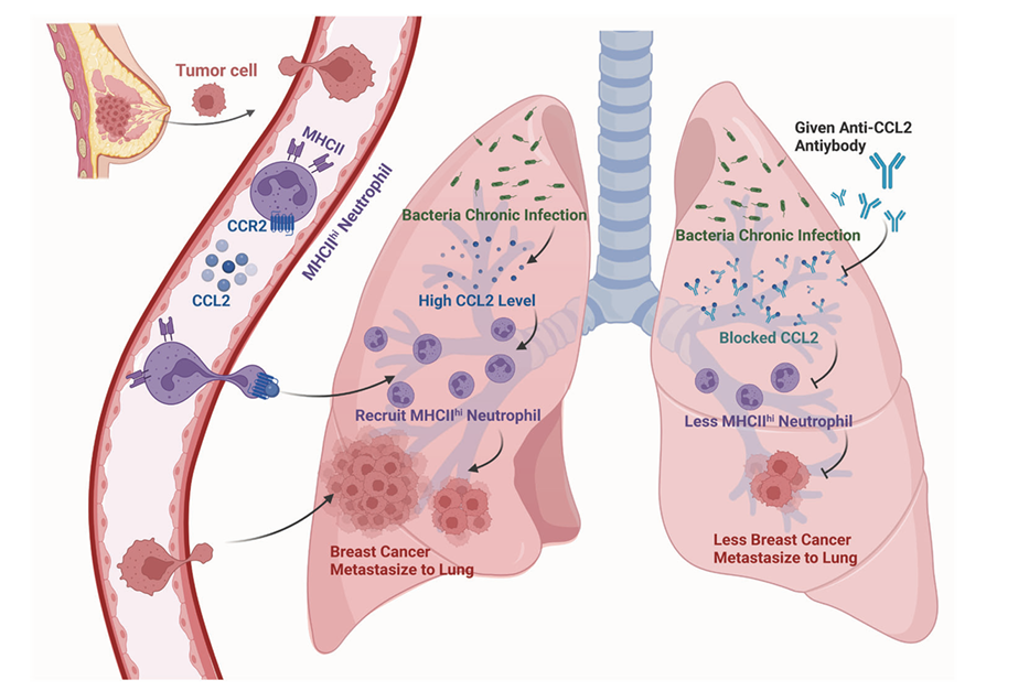
Fig. 2 Schematic representation of the proposed mechanism of chronic pulmonary bacterial infection-induced lung metastasis. Schematic was created with BioRender
03
Journal: Molecular Cancer
IF: 37.3
Title: Exosome-mediated miR-144-3p promotes ferroptosis to inhibit osteosarcoma proliferation, migration, and invasion through regulating ZEB1
Method: By transfection with LV or ADV, we knocked down or overexpressed the expression of miR-144-3p or ZEB1 in distinct groups of h143B cells and the significant transfection effectiveness was verified by RT-qPCR. (Fig. 3F, G).
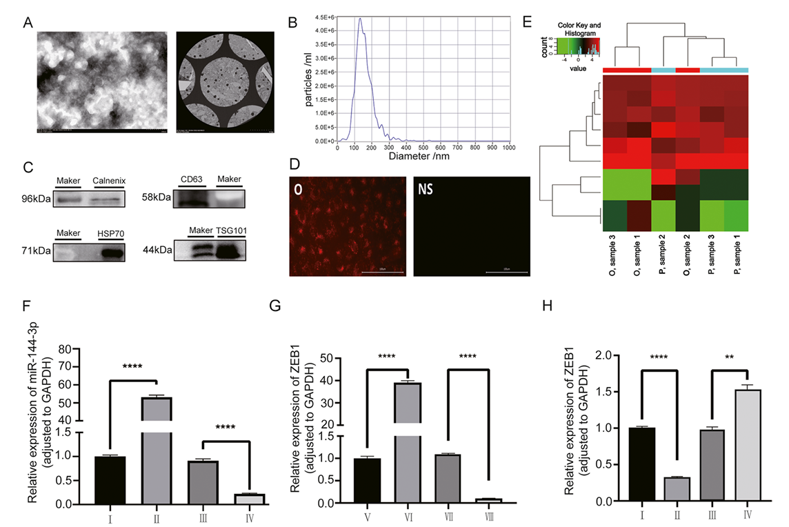
Fig. 3
04
Journal: Gastroenterology
IF: 29.4
Title: Pancreatic acinar cells-derived sphingosine-1-phosphate contributes to fibrosis of chronic pancreatitis via inducing autophagy and activation of pancreatic stellate cells
Method: A lentiviral vector containing the GFP-RFP-LC3B plasmid was constructed by HANBIO (Shanghai, China). PSCs transfected with GFP-RFP-LC3B were cultured in twenty-four-well plates with cover slips and then treated with CCK, PF-543, S1P, JTE013, RAP or CQ for 24 h respectively. After fixation with formalin (10%), the cover slips were mounted over a microscope slide. Confocal microscopy (Zeiss LSM 780, Germany) was used to examine changes in LC3B localization and expression. Images were obtained using confocal microscopy and analyzed with Zeiss confocal software.
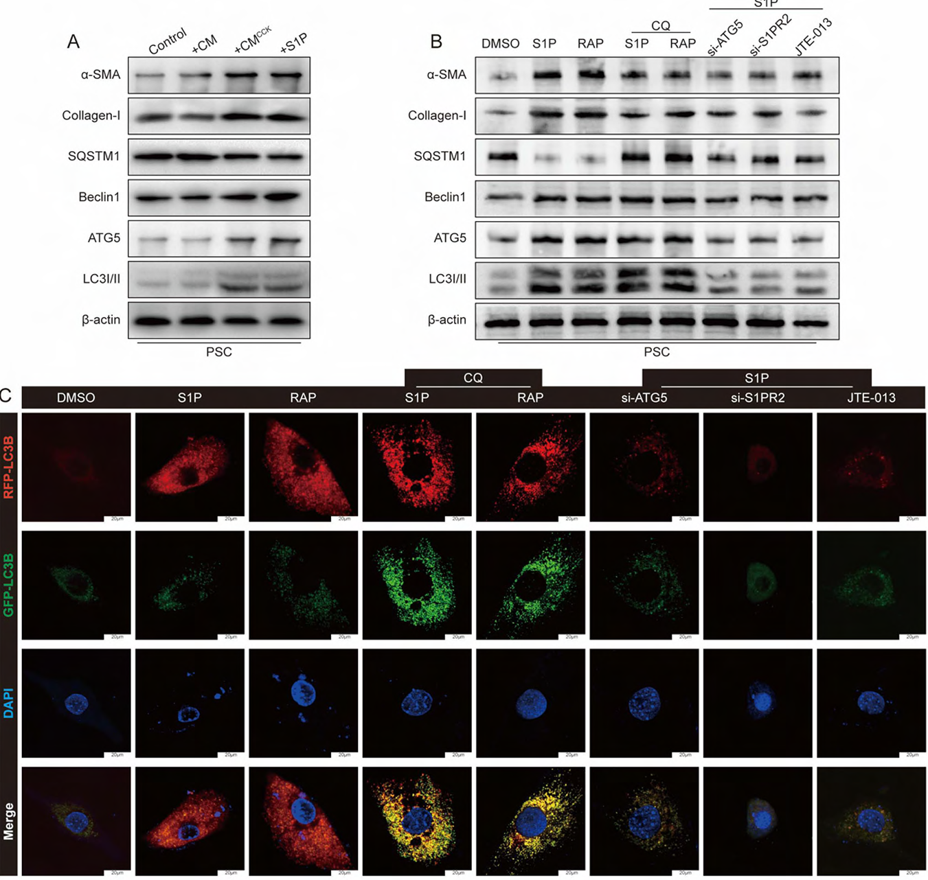
Fig. 4
05
Journal: Advanced Materials
IF: 29.4
Title: Satellite‐based On‐orbit Printing of 3d Tumor Models
Method: H460 and H460-Cis cells were stably transfected by lentiviral transfection with lentiviruses carrying GFP and RFP fluorescent protein genes (Hanbio, China) according to the commercial instructions. Briefly, cells were seeded in 6-well plates and grown to 30% confluence. Fresh medium with 2 μL of polybrene infection reagent and 50 μL of concentrated viral fluid was added, and the medium was changed after viral infection for 24 h. GFP- and RFP-labeled cells were visible under fluorescence microscopy 48–72 h after infection. The transfected cells were further enriched by puromycin treatment or flow cytometry sorting (Influx, BD Biosciences, USA).
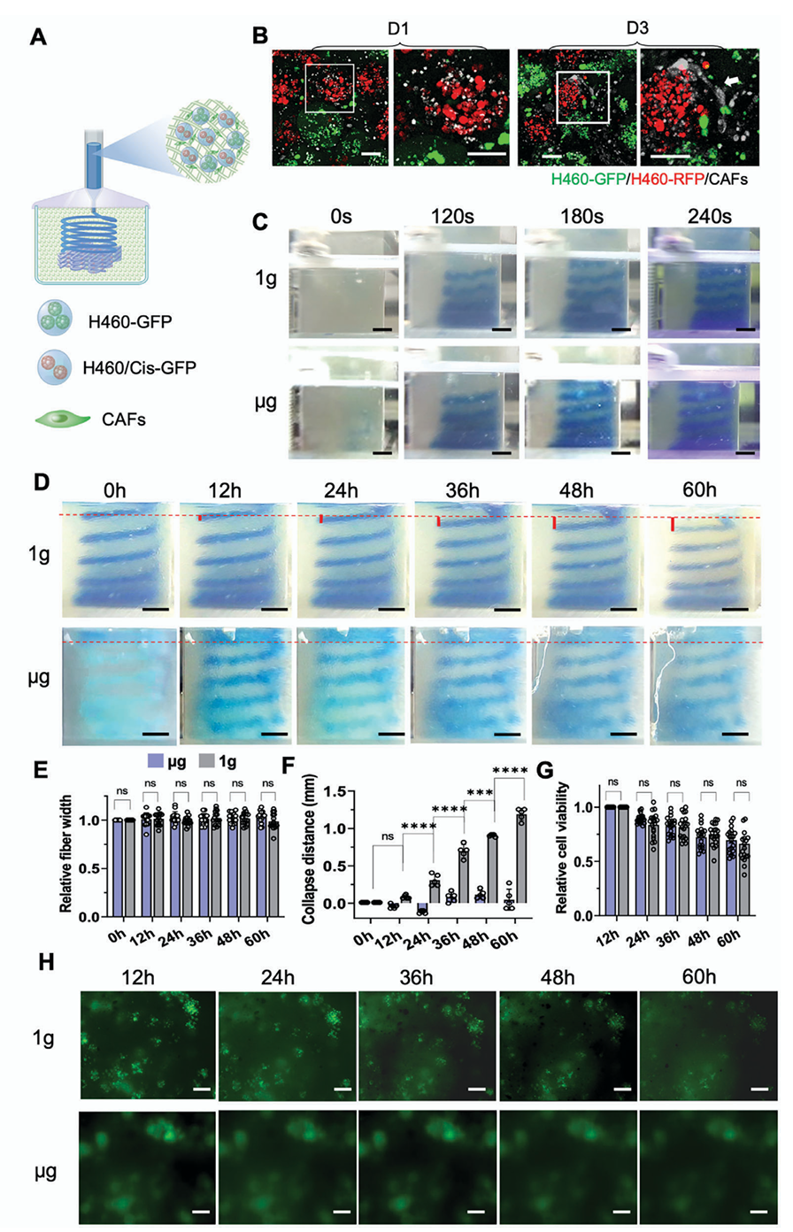
Fig. 5